

This is the story of a masked life-threatening ailment.
It was almost the closing time for the clinic after a busy day.
A 22 years old boy came with history of abdominal discomfort and dyspepsia.
The treating physician had advised Ultrasound of abdomen.
The boy had reported to the ARDISS IMAGING & DIAGNOSTIC CENTRE, Greater Noida, for the advised examination.
While performing the abdominal ultrasound, it was noted that the boy had accumulated little amount of fluid in his thorax, known as Pleural effusion in medical lexicon.
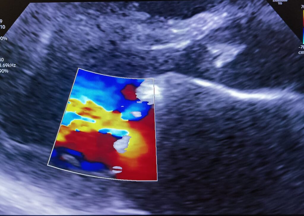
The suspicious finding prompted the senior experienced radiologist to place the probe in the subxiphoid region directed towards heart. The findings were alarming.
There was almost complete closure of one of the heart valves, namely the mitral valve, that is the connecting door between the two left sided cardiac chambers. The valve was thickened. Very little flow of blood was observed from one into the other chamber of the heart, namely from left atrium to left ventricle.
There was evidence of back pressure into the lungs that was causing stagnation of fluid inside the thorax. And now, why the boy was having intermittent little dry cough was convincingly explained by the findings. The retention of fluid in the lungs was causing the airway symptoms.
The abdominal symptoms were spurious. They were a distraction. The actual ailment was in the heart.
Ultrasound at the ARDISS IMAGING & DIAGNOSTIC CENTRE solved the physician’s diagnostic uncertainty.
Timely detection saved a lot of invaluable time and suffering for the patient.